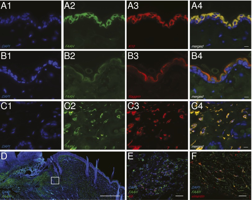
Fig. 1.
Localization of FAAH in adult mouse skin. (A–C) Representative double immunofluorescence images for FAAH, the keratinocyte markers cytokeratin-10 (K10) (A1–A4) and filaggrin (B1–B4), and the fibroblast marker vimentin (C1–C4) in intact skin. (Scale bars, 10 µm.) (D) Immunofluorescence localization of FAAH in wounded skin. The highlighted square is magnified in E and F, to show the presence of immunoreactive FAAH in keratinocytes (cytokeratin-5; E) and fibroblasts (vimentin; F). [Scale bars, 500 µm (D) and 20 µm (E and F)]. FAAH is shown in green, and all other markers in red; colocalization (A4, B4, and C4) is shown in yellow; nuclei are stained with DAPI (blue). An antibody selective for FAAH-1, the only FAAH isoform present in rodent tissues, was used in these experiments.

