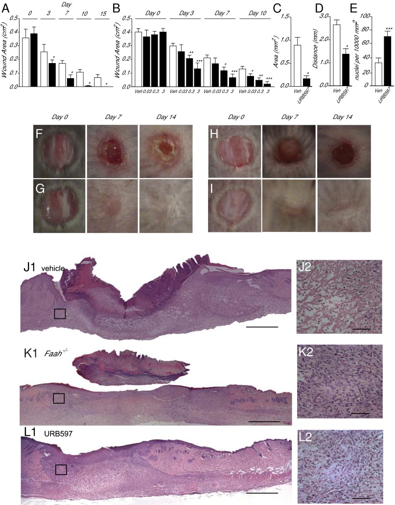
Fig. 2.
FAAH regulates healing of excisional skin wounds in mice. (A) Time course (days) of wound healing in wild-type mice (open bars) and Faah−/− mice (filled bars). (B) Time course of wound healing in wild-type mice treated with vehicle (open bars) or with various topical doses (%; wt/vol) of the FAAH inhibitor URB597 (filled bars). (C–E) Morphometric analyses showing the effects of vehicle (open bars) or URB597 (3%, wt/vol) (filled bars) on wound area (C), distance between migration tongues (D), and cell density in the dermis (E). (F–I) Representative images illustrating skin wound healing in drug-naïve wild-type mice (F), drug-naïve Faah−/− mice (G), and wild-type mice treated either with vehicle (H) or URB597 (3%, wt/vol; I). (J–L) Hematoxylin/eosin staining of skin sections from drug-naïve wild-type mice (J1), drug-naïve Faah−/− mice (K1), and wild-type mice treated with URB597 (3%, wt/vol; L1). The highlighted squares are magnified in J2, K2, and L2. [Scale bars, 500 µm (J1, K1, and L1) and 50 µm (J2, K2, and L2).] Skin sections were prepared 7 d after wounding. Data are expressed as mean ± SEM (n = 9). *P < 0.05; **P < 0.01; ***P < 0.001 (compared with vehicle or wild-type mice, two-tailed Student’s t test).

