Fig. 7.
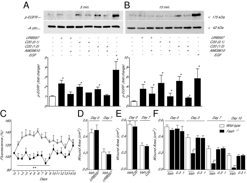
Signal transduction events initiated by FAAH-regulated NAT signaling in human keratinocytes. (A and B) Western blot analyses of primary cultures of human keratinocytes 5 min (A) and 15 min (B) after addition of the FAAH inhibitor URB597 or synthetic NAT(20:0) (C20 0.1 and 1 μM), in the absence or presence of the TRPV-1 antagonist AMG9810 (5 μM). (A, Upper, and B, Upper) Representative blots. (A, Lower, and B, Lower) Quantification of data from three independent experiments. EGF was used as positive control and β-actin as loading control. (C) Intracellular calcium levels (fluo-3 fluorescence) in human keratinocytes incubated with URB597 (1 μM; open circles) or URB597 plus TRPV-1 antagonist AMG9810 (5 μM; filled squares). Vehicle and AMG9810 had no effect on calcium levels when applied alone. (D) Effects of vehicle (open bars) or URB597 [3% (wt/vol); filled bars] on wound healing in Trpv1−/− mice. (E) Effects of vehicle (open bars) or NAT(20:0) [0.01% (wt/vol); filled bars] on wound healing in Trpv1−/− mice. In D and E, wound areas were measured 0 and 7 d after wounding. (F) Time-course of wound healing in wild-type mice treated with vehicle (open bars) and Faah−/− mice (filled bars) treated with vehicle or AMG9810 [0.3 or 1% (wt/vol)]. Data are expressed as mean± SEM (n = 3 for keratinocyte experiments, and n = 9 for in vivo experiments). *P < 0.05; **P < 0.01; ***P < 0.001 (compared with controls).

