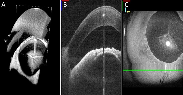
Figure 2.
Close-up view of the MI-OCT display options is demonstrated in A–C. Resident surgeons had a choice to view OCT display data of the surgical field during each maneuver in (A) a 3D view demonstrating the cornea (white arrow) and anterior chamber (red arrow), (B) cross-sectional B-scan view, or (C) as a surface volume projection.

