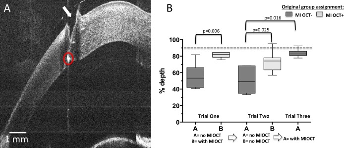
Figure 5.
The maneuvers of corneal laceration repair with goal depth of 90% corneal thickness. Fresh porcine corneas were used by MI-OCT− and MI-OCT+ groups. Fresh porcine corneas were used to construct vertical linear corneal laceration using a 15-blade scalpel. The corneal laceration repair with 10-0 nylon suture was performed by MI-OCT− and MI-OCT+ group residents. A representative cross-sectional B-scan was obtained orthogonal to the direction of needle pass and demonstrates a jagged break in corneal integrity consistent with corneal laceration (arrow) and a hyperreflective dot (circle) at goal depth of 90% corneal thickness (A). The average score of each resident was plotted, and the median depth scores compared across the groups using the Wilcoxon signed rank test, with relevant comparisons demonstrating statistical significance indicated at P < 0.05 (B).

