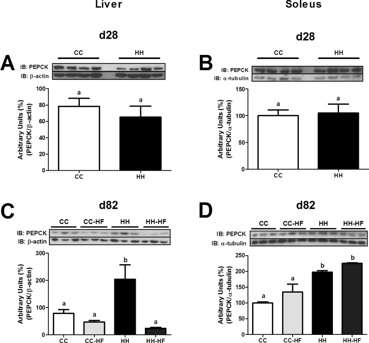
Fig 5. PEPCK content in liver and soleus.
Western blot analysis of PEPCK in liver at d28 (A) and d82 (C), in soleus at d28 (B) and d82 (D). For control of gel loading, membranes were reblotted with β-actin or α-tubulin. Data are means ± SEM (n = 3–8). Two-way ANOVA (C and D) or t test (A and B) was used. In all blots, at least three different litters were considered. Different letters indicate significant differences at p<0.05.

