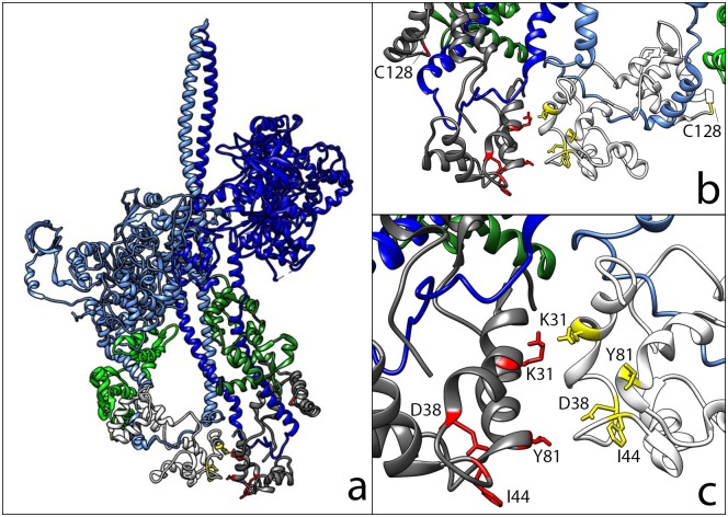
Fig 1. The structure of the tarantula IHM (3DTP) [9].
(A) The myosin heavy chain of the blocked head is light blue, and that of the free head is dark blue. The essential light chains are in light green and green, and the RLCs are in white and grey in the lower part of the structure. (B) The structure of the RLC domain as seen from the back of panel a, enlarged. Mutants are in yellow and red, C128 residues are highlighted. The first 50 residues in the N-terminus of the RLCs are not shown since they are very mobile and not present in the mouse RLC. (C) A closer view of the two RLC structures, as seen in panel b, with the mutants highlighted.

