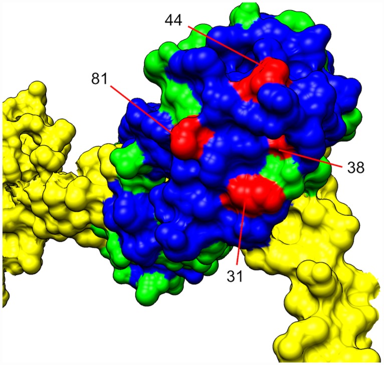
Fig 3. The structures of the N-terminal lobe of the RLC along with a portion of the heavy chain (in yellow) model are shown (picture adjusted from the 3J04 model of chicken smooth muscle myosin and RLC [35]).

The non-conserved residues of the RLC are shown in green, the conserved residues are in blue, and 4 of the mutated residues are in cyan. The mutants, K31C, D38C, I44C and T81C are identified by their positions in the mouse construct used in our experiments. Conserved residues were identified by the chicken smooth muscle RLC and mouse skeletal muscle RLC alignment (S6 Fig).
