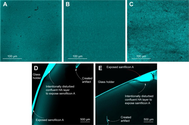
Figure 3.
CLSM images.
Notes: Balafilcon A (A), senofilcon A (B), and samfilcon A (C) incubated overnight with 0.1% (w/v) HA solution, then stained with safranin; images of senofilcon A (D), and samfilcon A (E) after scratching the sorbed HA layer with forceps. HA on balafilcon A could not be imaged by this method, because the stained lens did not sufficiently adhere to the glass slide. A–C Captured using a 20× microscope objective and 2× confocal magnification; D–E were captured using a 4× microscope objective.
Abbreviations: CLSM, confocal laser-scanning microscopy; HA, hyaluronic acid.

