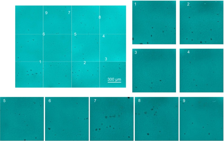
Figure 7.
CLSM image of HA distribution over a large senofilcon A lens area.
Notes: The master image of the stained HA network distributed over the entire lens area was prepared by stitching sequential images recorded using a 10× objective. Randomly chosen areas were examined using 20× magnification to visualize the HA-network details. The beads (fiducial marks) were used to localize the image position on the master image.
Abbreviations: CLSM, confocal laser-scanning microscopy; HA, hyaluronic acid.

