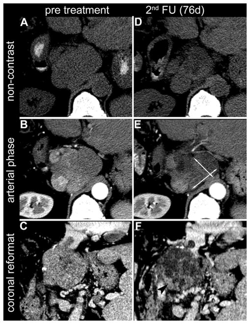
Figure 3.

Axial slices of non-contrast CT (A, D) and arterial phase CT (B, E) of a 66-year-old male with HCC in liver segment I. In the 2nd follow-up CT (B,E), 76 days after locoregional treatment, the bidimensional measurement of the remaining enhancing tumour tissue (EASLmeas; arrows in E) indicated a decrease of 20% as compared to the baseline scan and therefore resulted in a response evaluation of SD (stable disease). By visual estimation (EASLest), the same reader judged the reduction in enhancing tumour tissue to be 70%, resulting in a response evaluation of PR (partial response). This visual estimation of reduced enhancing tumour tissue (arrowhead in F) is supported by the coronal reconstructions of the tumour shown on the baseline CT (C) and at 2nd follow up (F).
