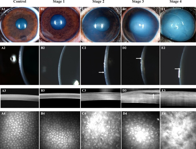
Figure 1.
Corneal structure and cellular morphology as assessed by color photography, digital slit-lamp biomicroscopy, FD-OCT, and IVCM dramatically differed between control and CED-affected BTs with varying stages of severity. An 8-year-old castrated male BT with a clear cornea (A1, A2), normal corneal thickness, and lamellar arrangement of the collagen fibrils (A3), and regular, hexagonal arrangement of corneal endothelium (A4); this dog was categorized as a control. A 6-year-old castrated male BT with a clear cornea (B1, B2), normal corneal thickness, and lamellar arrangement of the collagen fibrils (B3) but moderate pleomorphism and polymegathism of the corneal endothelium (B4); this dog was the son of a CED-affected dog and was categorized as stage 1. An 11-year-old spayed female BT with mild edema in the temporal paraxial and perilimbal cornea (C1, C2) with microcystic bullae in the anterior stroma (C2, white arrow), increased corneal thickness (C3), and moderate pleomorphism and marked polymegathism of the corneal endothelium (C4); this dog was categorized as stage 2. An 11-year-old spayed female BT with moderate edema in the central, temporal paraxial and perilimbal cornea (D1, D2), increased corneal thickness with an axial anterior stromal bulla (D2, D3, white arrows) and marked pleomorphism and marked polymegathism of the corneal endothelium (D4); this dog was categorized as stage 3. A 14-year-old spayed female BT with marked, diffuse corneal edema (E1, E2), microcystic bullae in the anterior stroma (E2, white arrow), marked increase in corneal thickness (E3), and loss of the orderly arrangement of collagen fibrils within the anterior stroma (E3, E4), such that the corneal endothelium and keratocytes could not be visualized (E4); this dog was categorized as stage 4.

