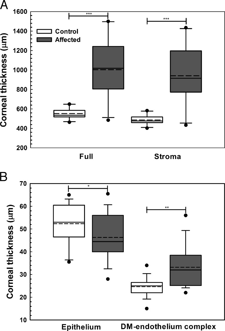
Figure 3.
Central corneal full, stromal, epithelial, and DM-endothelium complex thickness significantly differed between CED affected and control (unaffected) dogs as measured by FD-OCT. Central corneal thickness was significantly greater in CED-affected versus unaffected dogs primarily due to a significant increase in stromal thickness in CED-affected versus unaffected dogs (A). Epithelial thickness in the central cornea was significantly less in dogs with CED versus control dogs (B). In the central cornea, DM-endothelium complex thickness was significantly greater in dogs with CED versus control dogs (B). Box plots depict median (solid line), mean (dashed line), 25th, and 75th percentiles, while whiskers show 10th and 90th percentiles; black circles indicate outliers. The P values were determined by a Mann-Whitney rank sum test (CCT, stroma, and DM-endothelium complex) or Student's t-test (epithelium), *P < 0.05, **P < 0.01, and ***P < 0.001.

