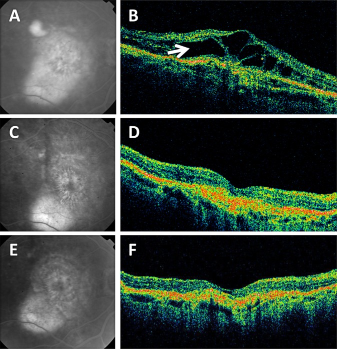
Figure 2.
Late fluorescein angiographic (FA) images and time-domain optical coherence tomography (OCT) diagonal B-scans from a patient treated with intravitreal ranibizumab in the phase II study, then followed in the extension study with observation and no additional retreatments with ranibizumab.17 (A, C, E) Late FA images. (B, D, F) Diagonal OCT B-scans. (A) Baseline late FA image showing diffuse leakage. Visual acuity (VA) was 20/125. (B) Corresponding baseline OCT image showed cystic macular edema (arrow). (C) After eight injections over 140 days, the FA leakage had significantly diminished, and VA improved to 20/80. (D) Corresponding OCT B-scan showed resolution of the macular edema. (E) After an additional 22 months without any ranibizumab injections, VA improved to 20/32 and there was no evidence FA leakage. (F) Corresponding OCT B-scan showed a persistently dry macula.

