
Figure 2.
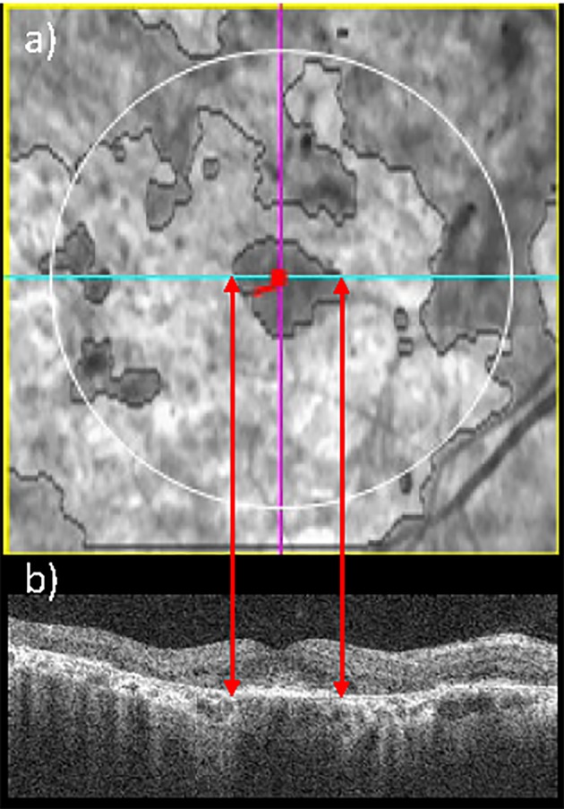
Example of en face SD-OCT, which enables to identify a STGD1 patient with foveal sparing. En face SD-OCT fundus image (a): the red dot represents the fovea (automatically detected) and the light blue line indicates the corresponding B-scan. Spectral-domain OCT B-scan corresponding to the fovea (b) highlights that the fovea is not affected by RPE lesion.