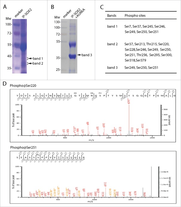
Figure 3.
SOX2 was multiple phosphorylated in M phase. (A) SOX2 protein was separated from mitotic PA-1 cells using SOX2 specific antibody. SDS-PAGE separated proteins were stained and extracted for mass spectrometry analysis. (B) Coomassie brilliant blue staining of SOX2 protein in AURKA kinase assay. PA-1 cells were incubated with VX680 (0.5 μM) for 5 hours and then immunoprecipitated using SOX2 specific antibody. SOX2 proteins were subjected to in vitro kinase reaction with AURKA. Reaction products were SDS-PAGE separated and analyzed by mass spectrometry. (C) Confirmed phosphorylation sites in SOX2 proteins from band 1, band 2 in (A), and band 3 in (B). (D) Two representative spectrums of SOX2 phosphorylations on sites Ser220 and Ser251.

