
Figure 2.
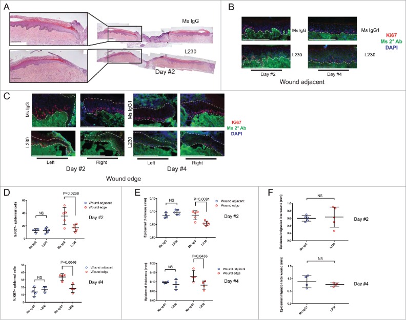
Integrin αv is necessary for proliferation during wound healing of human skin in vivo. (A) Representative Hematoxylin and Eosin (H&E) stains for wounds from mice treated with a control mouse IgG antibody or an L230 antibody. Wounds were harvested 2 days after wounding. B,C. Representative images of Ki67 and mouse secondary antibody labeling of wound adjacent tissue (B) or wound edge tissue (C). Dotted lines indicate the basement membrane zone. (D, E) Quantification of %Ki67+ epidermal cells (D) or epidermal thickness (E) at the wound edge and in wound adjacent tissue. Wound edge was considered within 0.25mm of wound. (F) Quantification of epidermal migration into the wound in mm. N = 4 mice per group for day 2 and 5 mice per group for day 4. NS = not statistically significant. P values were calculated using a student's t-test. Scale bar = 200 µm.