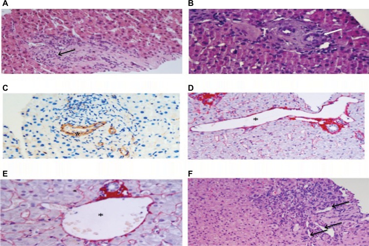
Figure 1.
Histologic features of the portal tracts in INCPH.
Notes: (A) Obliterative venopathy: portal vein with a reduced lumen (arrow) within a fibrotic portal tract; hematoxylin and eosin stain, original magnification ×10. (B) Obliterative venopathy: a fibrotic portal tract (white arrow) with a rounded contour and a small portal branch showing a thickened wall (arterialization): hematoxylin and eosin stain, original magnification ×20 (C) Obliterative venopathy: immunostain with antismooth muscle actin antibodies highlights smooth muscle cells hyperplasia in portal vein branch arterialization (asterisk); original magnification ×20. (D) Paraportal shunt: dilated, thin-walled portal vascular channel herniating into the surrounding parenchyma (asterisk); picrosirius red stain, original magnification ×10. (E) Marked portal vein dilation: the enlarged portal branch (asterisk) is at least three times greater than the size of the bile duct; picrosirius red stain, original magnification ×20. (F) Increased number of portal vascular channels (arrows); hematoxylin and eosin stain, original magnification ×10.
Abbreviation: INCPH, idiopathic noncirrhotic portal hypertension.

