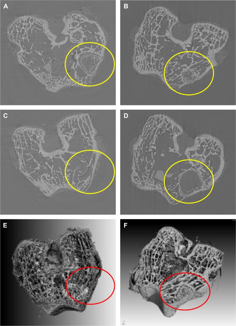
Figure 8.
SRmCT images of central virtual slice of MWC and WP scaffolds and cross-section of 3D reconstruction SRmCT images of MWC and WP scaffolds.
Notes: SRmCT images of central virtual slice of MWC scaffolds implanted into the femoral defects (yellow circle) of rabbits for 4 (A), 8 (B), and 12 (C) weeks and WP scaffolds implantation for 12 (D) weeks. Cross-section of 3D reconstruction SRmCT images of MWC scaffolds (E) and WP (F) scaffolds implanted into the femoral defects of rabbits for 12 weeks, and the red circles describe the defects.
Abbreviations: 3D, three dimensional; MWC, nano magnesium phosphate/wheat protein composite; SRmCT, synchrotron radiation microcomputed tomography; WP, wheat protein.

