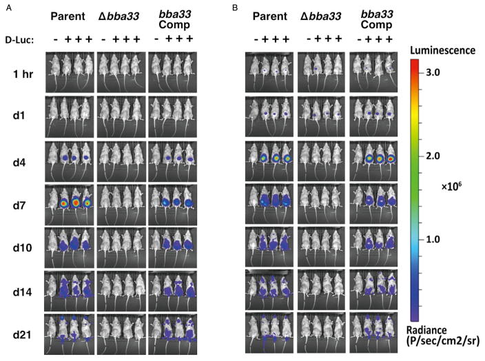
Fig. 5.
Temporal and spatial tracking of Borrelia burgdorferi strains following infection with 103 and 105 spirochetes. C3H mice were infected with the parent (Parent; ML23 pBBE22luc), the Δbba33 mutant (Δbba33; HZ001 pBBE22luc) or the bba33 complemented strain (bba33 Com., HZ001 pHZ300) at a dose of 103 (A) and 105 (B). Mice denoted with ‘+’ were treated with D-luciferin and imaged at the times listed on the left. For each image shown, the mouse on the far left (denoted as ‘−’) was infected with B. burgdorferi but did not receive D-luciferin to serve as a background control. All images were normalized to the same scale (shown on the right).

