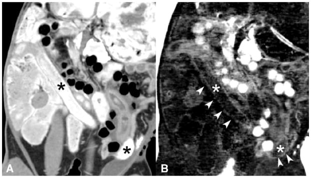
Figure 1.
Simultaneous enteric bismuth (Bi) contrast and intravenous iodine contrast in a rabbit model. Conventional CT image (A) shows bright enteric bismuth contrast in the bowel lumen (*) that limits visualization of the curvilinear bowel wall enhanced by intravenous iodine. The DECT iodine density map (B) shows partial subtraction of bismuth enteric contrast (*), but the curvilinear bowel wall enhancement from iodine (arrowheads) is not well seen. The DECT iodine map image was not significantly preferred over the co-registered conventional CT image in regards to small bowel wall visualization. Note also that air appears bright on the iodine/bismuth material decomposition (B) due to a current software-associated air artifact, further detailed in the methods section.

