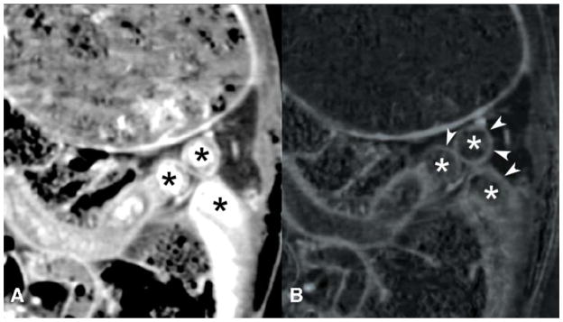
Figure 3.
Simultaneous enteric tantalum (Ta) contrast and intravenous iodine contrast in a rabbit model. Conventional CT image (A) shows bright enteric tantalum contrast in the bowel lumen (*) that limits visualization of the curvilinear bowel wall enhanced by intravenous iodine. The iodine density image map (B) from DECT shows improved small bowel wall (arrowheads) visualization compared to the co-registered image from the conventional CT (A). * = bowel lumen, which is bright in the conventional CT image due to tantalum contrast, but is dark in the iodine density image where the tantalum contrast is subtracted.

