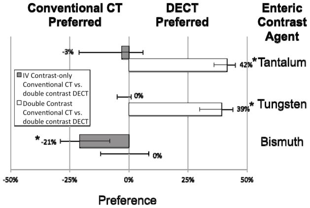
Figure 5.
Pairwise comparison of small bowel wall visualization at conventional CT versus iodine density maps from double-contrast enhanced DECT with enteric bismuth (or tungsten or tantalum) contrast and intravenous iodine contrast in rabbits. Scored median improvement in small bowel wall visualization is shown by bar graph for comparing intravenous-contrast enhanced conventional CT (to the left of the bold line) versus DECT iodine maps (to the right of the bold line) in the case of tantalum, tungsten, and bismuth contrast respectively. Whiskers denote the 95% confidence interval. * = p < 0.001

