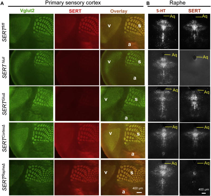
Fig. 2. SERTGluΔ but not SERTCortexΔ or SERTRapheΔ abolishes SERT expression in primary sensory cortices.

A. SERT (red) and Vglut2 (green) double immunostaining of tangential sections of primary visual (v), auditory (a) and somatosensory barrel (s) cortices in P7 mutant and control SERTfl/fl littermate mice. SERT staining in Vglut2+ patches in SERTCortexΔ mice was indistinguishable from the SERTfl/fl control or SERTRapheΔ mice, whereas the SERT staining in SERTGluΔ mice was diminished as in SERTNull mice. B. SERT and 5-HT immunostaining (both grayscale) of adjacent coronal sections of raphe nuclei from P7 mice. SERT staining was abolished in SERTNull, diminished in SERTRapheΔ, but unaffected in SERTGluΔ and SERTCortexΔ mice. Aq, aqueduct. Some images for SERTNull, SERTRapheΔ and SERTGluΔ brain sections are reproduced from a previous study (Chen et al., 2015) for comparison of SERT immunostaining patterns in SERTCortexΔ brain sections. (For interpretation of the references to colour in this figure legend, the reader is referred to the web version of this article.)
