
Fig. 4. SERT knockout in TCAs not in cortical neurons disrupts barrel map formation.
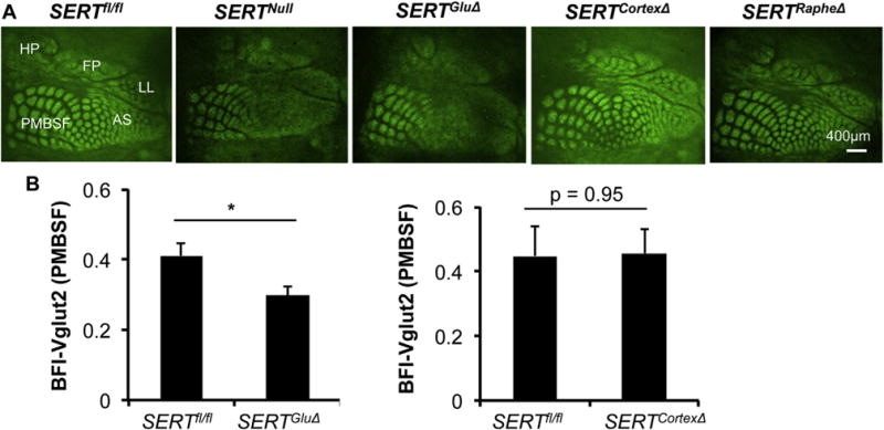
A. TCA patterning at L4 visualized by Vglut2 immunostaining of tangential sections through the somatosensory cortex of P7 mice. In control SERTfl/fl mice, Vglut2+ TCAs corresponding to distinct body parts display arrayed patches in five indicated reception subfields. In SERTGluΔ and SERTNull mice, the areas for the subfields were preserved, but no discernible Vglut2+ patterning in the subfields corresponding to anterior snout (AS), lower lip (LL), forepaw (FP) and hindpaw (HP), and Vglut2+ patches in PMBSF corresponding to whiskers were more blurred although the patches were preserved. Vglut2+ patterning in all the subfields in SERTCortexΔ mice was indistinguishable from that seen in SERTRapheΔ mice and control littermate mice. Images for SERTNull, SERTRapheΔ and SERTGluΔ cortices are reproduced from a previous study (Chen et al., 2015) for comparison of Vglut2 immunostaining and TCA patterning in SERTCortexΔ barrel cortex. B. Evaluation of Vglut2+ TCA patterning in P7 PMBSF, by calculating BFI-Vglut2. Mutant and control littermate mouse samples were stained in parallel. N = 5 each for SERTGluΔ and SERTfl/fl littermate mice; N = 3 each for SERTCortexΔ and SERTfl/fl littermate mice, mean ± SEM, *, p < 0.05, Student’s t-test.