
Fig. 5. SERT knockout in TCAs not in cortical neurons disrupts cytoarchitectural organization of Satb2+ neurons in the primary somatosensory cortex.
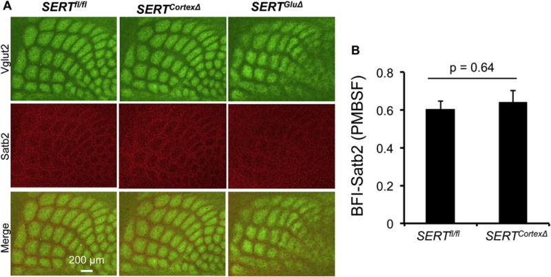
A. Tangential sections of P7 PMBSF of the somatosensory cortex labeled by double immunostaining of Vglut2+ TCAs (green) and Satb2+ cell nuclei (red). In control littermate mice, Satb2+ cells form a ring structure surrounding Vglut2+ TCA patches. This Satb2+ topographic patterning was preserved in SERTCortexΔ mice. B. Evaluation of Satb2+ neuron patterning in the barrel cortex based on BFI determination using Satb2 immunohistochemistry in P7 PMBSF. N = 4, mean ± SEM, Student’s t-test. (For interpretation of the references to colour in this figure legend, the reader is referred to the web version of this article.)