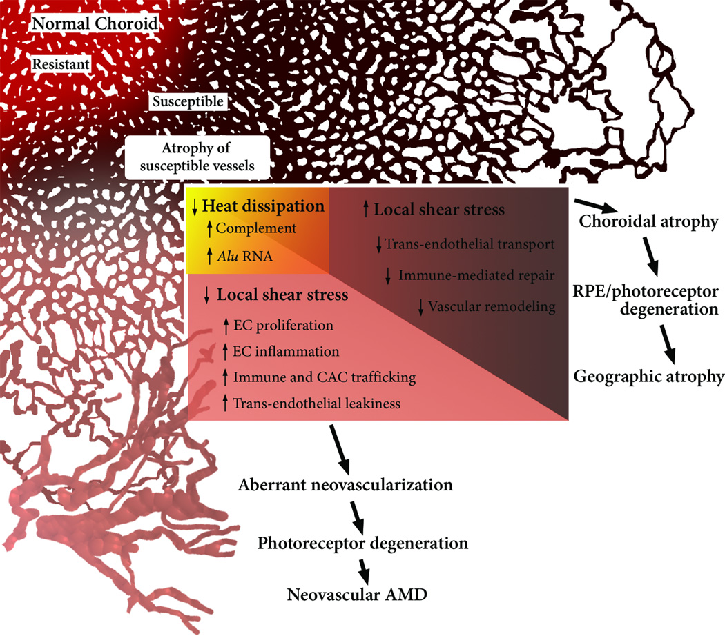
Key Figure, Figure 4. A Revised Hemodynamic Model of AMD.
The model posits that the normal, healthy choriocapillaris consists of resistant (red) and susceptible (brown) areas, based on the local hemodynamic environment. Relevant factors in addition to those mentioned by Friedman include distance from the terminal arteriole, density of arterioles and venules, as well as choriocapillaris thickness and density. With aging, susceptible areas may eventually adopt an atrophic phenotype (beige), which alters local hemodynamics. By virtue of decreased CC density, atrophied areas lose heat-radiating capacity, thereby increasing inflammatory mediators such as complement and Alu RNA. In areas of the CC, atrophy induces elevated shear stress in adjacent capillaries, rendering the choroid ill-suited to remove retinal waste and incapable of vascular repair. This ultimately leads to GA by choroidal RPE and retinal atrophy. Other areas of the CC experience reduced shear stress, thereby promoting an ‘activated’ endothelial phenotype. The angiogenic and inflammatory influences of low shear stress and hyperthermia of the RPE conspire to produce a choroidal neovascular membrane.

