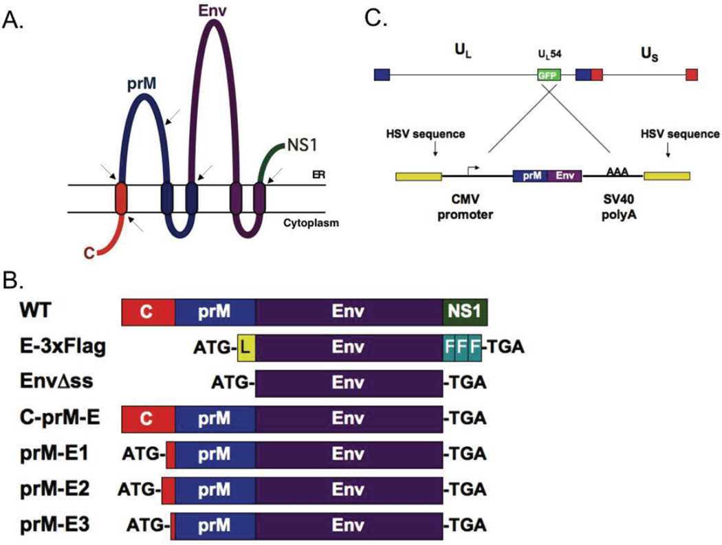
Figure 1. Diagrams of the constructs used in this study.
A. Graphical representation of the membrane orientation of the structural proteins in WNV including the initial portion of the nonstructural NS1 protein. Transmembrane regions are denoted by ovals passing through the lipid bilayer. Arrows indicate where cellular or viral proteases cleave the polyprotein to free the individual proteins: capsid (C), premembrane (prM), and envelope (Env). B. Comparison of the various plasmid constructs to the wildtype (WT) structural protein coding sequences. All plasmids used the CMV promoter/enhancer to drive transcription. The addition of start (ATG) and stop (TGA) codons are indicated. E-3xFlag contains the preprotrypsin leader sequence (L) and three sequential FLAG epitope tags (F). EnvΔss expresses the E protein lacking C and prM sequences. C-prM-E contains the WNV coding sequences for C-prM-Env-NS1. prM-E1-3 contain varying amino acid residues of C as described in Table 2. (C) Top: the d106 genome with the location of the UL54 (ICP27) region containing the GFP transgene indicated. Bottom: the pd27-WNV plasmid used to generate the recombinant d106-WNV vaccine vector. The crossed lines show the homologous recombination event between the reversed plasmid sequences with the d106 genome that generated the d106-WNV recombinant virus.

