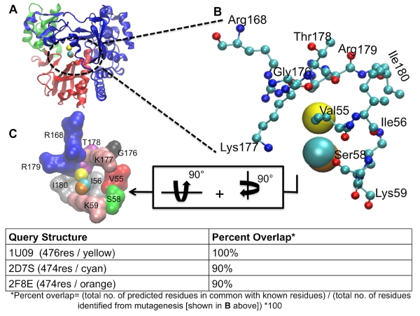
Figure 3.
LISE accurately predicts the site of a known allosteric inhibitor of the FMDV polymerase. Panel A shows the rear of the enzyme with the LISE predicted spheres from the 3 query structures in van der Waals (VDW) representation (1U09 in yellow, 2F8E in orange and 2D7S in cyan). Panel B is a magnified view of A showing only the residues (in CPK representation) that have been biochemically established to be critical for ligand binding at this site and the LISE predicted spheres (results for query structure 1U09 in yellow, 2F8E in orange and 2D7S in cyan). Panel C displays the residues making up this site depicted in surface representation after 90° rotations about both the y and x axes. Each residue is separately colored and labeled.

