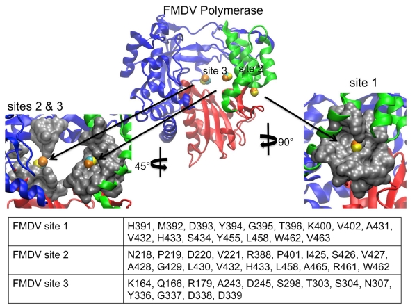
Figure 9.
LISE predicted sites in the FMDV polymerase. The center image depicts the FMDV enzyme LISE predicted spheres in the palm and thumb domains (1U09 in yellow, 2F8E in orange and 2D7S in cyan). The left image is a magnified surface view of both of the predicted sites 2 and 3 located in the palm domain that share structural similarities with the HCV NNI-3 pocket. The right image is an enlarged surface view of the predicted site 1 which is structurally similar to the HCV NNI-2 pocket. The table lists the consensus residues making up each LISE predicted site.

