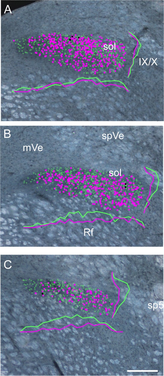
Fig. 4.

Plots of the distribution of rNST GAD65 and PHOX2b neurons from a different GAD65-Cre × tdTomato mouse than shown in Fig. 3. GAD65/tdTomato and PHOX2b neurons are depicted with magenta and green symbols, respectively. The few double-labeled neurons are shown with black symbols. Plots are superimposed upon darkfield photomicrographs of the same sections. A–C show successively more rostral levels of the nucleus. Frequency polygrams for the distributions of GAD65 and PHOX2b neurons along the mediolateral and dorsoventral axes appear in matching solid lines for each panel. GAD65 neurons are sparse at the most medial pole of the nucleus, but this region is occupied by PHOX2b-stained nuclei. We observed that these medial PHOX2b nuclei had a relatively large diameter, suggesting that they are associated with preganglionic parasympathetic neurons (Contreras et al. 1980; Kang et al. 2007). Otherwise, GAD65 neurons are distributed throughout the nucleus. Note that although this case showed a tendency for a higher proportion of GAD65 neurons caudally, this was not consistent in the other cases inspected. IX/X, incoming fibers of the glossopharyngeal and vagus nerves; mVe, medial vestibular nucleus; spVe, spinal vestibular nucleus; sp5, spinal trigeminal tract; sol, solitary tract; Rf, reticular formation. Scale bar, 200 μm.
