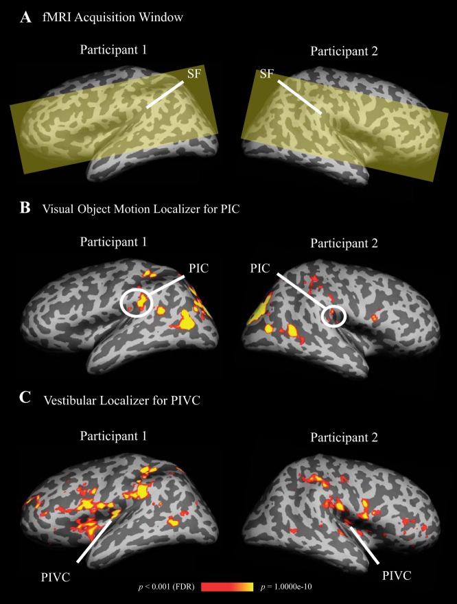
Fig. 2.
Definition of areas posterior insular cortex (PIC) and parieto-insular vestibular cortex (PIVC) by means of activations in visual object motion and caloric stimulation, shown on inflated left and right hemispheres of two sample participants. PIC and PIVC were also activated in the other hemisphere of the shown participants, however, not at the conservative threshold (p < 0.001, false-discovery-rate corrected), used here for displaying purpose. A: the functional MRI acquisition window (yellow shading) was restricted to the region surrounding the Sylvian fissure (SF). B: significantly stronger activation in PIC during visual object motion compared with baseline (=static objects). Color-coding ranges from red to yellow and indicates increasing levels of statistical significance (scale bar shown at bottom). In both sample participants PIC was split in anterior and posterior clusters. C: significantly stronger activation in PIVC during caloric stimulation compared with baseline (=warm in both ears). Color coding as in B.

