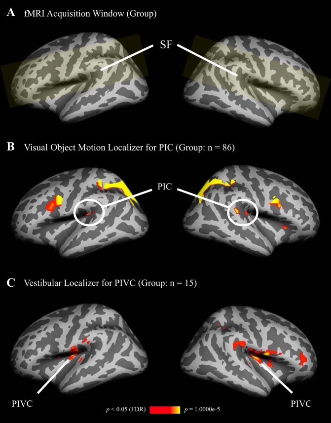
Fig. 3.
Average locations of areas PIC and PIVC, revealed by random-effects group analyses, and shown on inflated left and right hemispheres of the Freesurfer template brain (color coding as in Fig. 2). A: common acquisition window (yellow shading) was restricted to the region surrounding the SF, following the data acquisition window for the current study (see Fig. 2A). B: significantly stronger activation in PIC during visual object motion compared with static objects. Since area PIC is small and varies substantially in location between participants, 86 subjects, who completed the object motion localizer between the years 2011 and 2015 for various studies in our laboratory, were included to reveal activation in PIC on the group level. One anterior and one posterior cluster for PIC emerged (see also Fig. 2B). C: significantly stronger activation in PIVC during caloric stimulation compared with baseline (n = 15).

