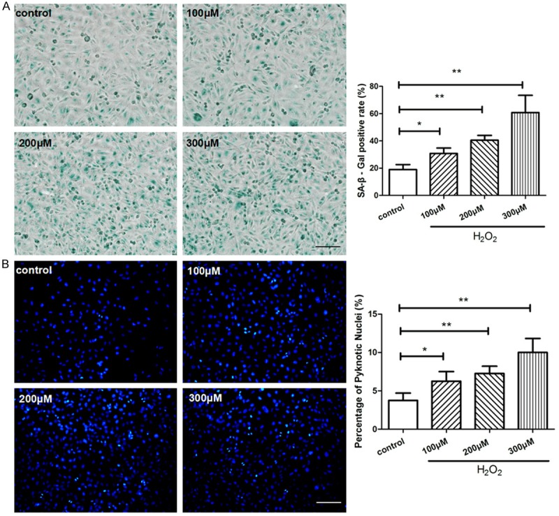
Figure 1.

H2O2 caused HUVECs senescence and apoptosis. HUVECs were treated with 0, 100, 200, 300 μM of H2O2 respectively for 4 hours and then cultured for another 24 hours. (A) SA-β-gal staining was performed to detect senescent cells (blue color), the ratio of SA-β-gal positive cells was calculated in each group. (B) The apoptosis assay was performed by Hoechst 33258 staining, pyknotic and brighter nuclei is the feature of apoptosis. Quantitative analysis was represented as the apoptotic cells in the total cells per field. Values are mean ± SEM; n = 4 in each group, N.S. means no significant difference, *means P<0.05, **means P<0.01, v.s. control group. Scale bar indicated 100 μm in (A) and 50 μm in (B). One-way ANOVA (Bonferroni post hoc test) was used.
