
Figure 3.
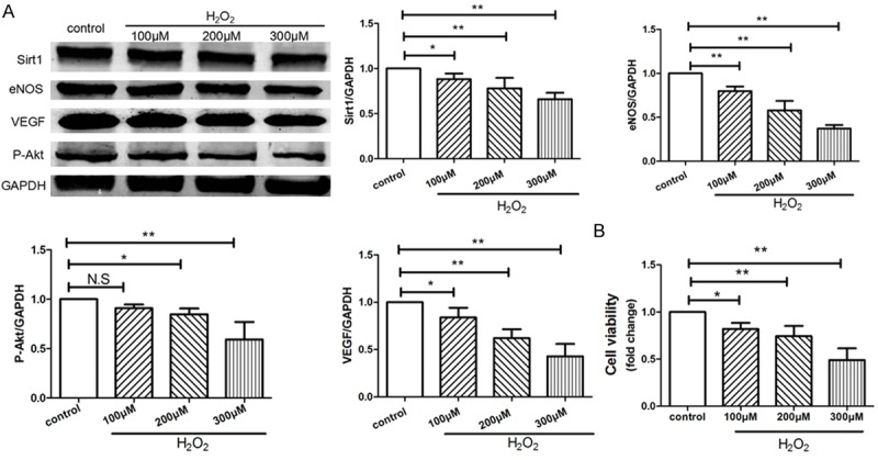
H2O2 inhibited angiogenesis and proliferation of HVUECs. HUVECs were treated with 0, 100, 200, 300 μM of H2O2 respectively for 4 hours and then cultured for another 24 hours. A: The protein expressions of SIRT1, VEGF, phospho-Akt, and eNOS in HUVECs were examined by Western blotting, data were represented as fold of control. B: The proliferation was analyzed by Cell Count Kit-8 (CCK-8) as indicated. Values are mean ± SEM; n = 4, N.S. means no significant difference, *means P<0.05, **means P<0.01, vs. control group. One-way ANOVA (Bonferroni post hoc test) was used.