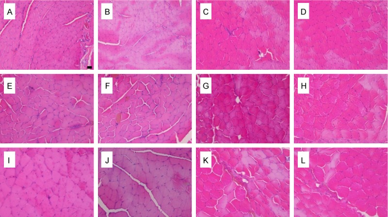
Figure 6.

Light microscopy images of transverse sections of the peroneal-innervated and tibial-innervated muscles, A-D, E-H, I-L: Images at 2, 4 and 8 months after nerve repair, respectively; A, E and I: Peroneal-innervated muscles on the operated side; B, F and J: Tibial-innervated muscles on the operated side; C, G and K: Normal peroneal-innervated muscles; D, H and L: Normal tibial-innervated muscles. (Scale bar=10 uM).
