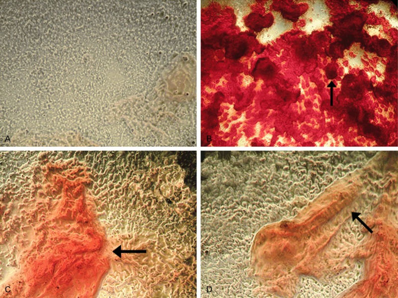
Figure 3.

Alizarin Red S staining of calcium accumulation in BMSCs cultured with EGB under light microscope. (A) Control group with no EGB; (B) Cells incubated with 150 μg/mL EGB; (C) Cells incubated with 50 μg/mL EGB; (D) Cells incubated with 400 μg/mL EGB. The calcium deposits are indicated by black arrows (X40). The highest calcium accumulation was observed at 150 μg/mL EGB.
