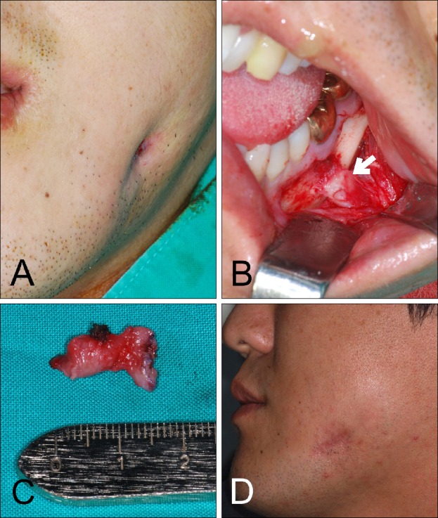
Fig. 4. Odontogenic cutaneous fistula of the mandibular body region. (A) Extraoral photo; showing dimpling on the left mandibular body region. (B) The odontogenic cutaneous fistula was observed by orientating the root apex (#36; left mandibular first molar) to overlying cortical plates and muscular attachments. The fistula tract is indicated by a white arrow. (C) Resected fistula tract. (D) Postoperative photo showing diminished dimpling.

