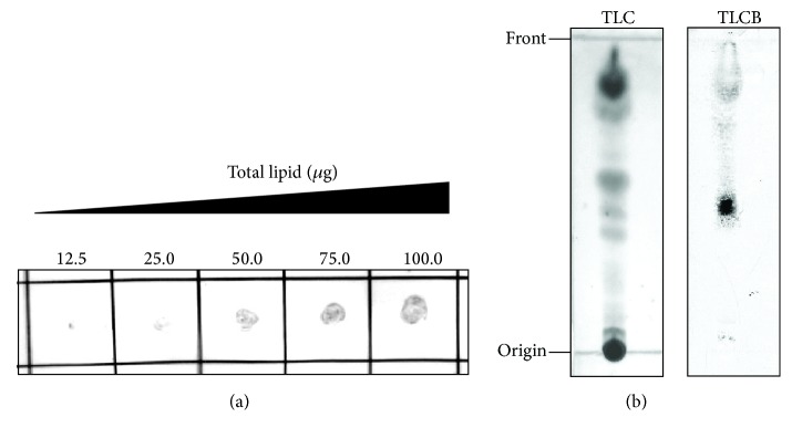
Figure 2.
Interaction between lipids of M. avium and HDL. (a) The lipids extracted from M. avium were spotted in the indicated amount (12.5–100.0 μg) on a nitrocellulose membrane. The membrane was then incubated with HDL (50 μg protein/mL), and HDL bound to the lipids was visualized using an anti-apoA-I antibody. (b) TLC plates with the spotted lipids (150 μg each sample) extracted from M. avium were developed using CHCl3/CH3OH (95 : 5, v/v). One plate was treated with a 20% H2SO4 solution to visualize the fractionated lipids (TLC). The fractionated lipids on the other plate were thermally transferred onto a PVDF membrane, which was then incubated with HDL (83 μg protein/mL). The lipid that bound to HDL was visualized using an anti-apoA-I antibody (TLCB).

