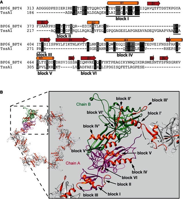
Figure 5. Secondary‐structure‐weighted sequence alignment of TssA1 with gp6.

- Protein sequences were extracted from UniProt accession number BP06_BPT4 for enterobacteria phage T4 (gp6; bacteriophage T4) and the Pseudomonas Genome Database (TssA1; reference strain PAO1). Conserved positions are shown in black and grey background. The secondary structural elements corresponding to the 3D structure of gp6 (PDB code 3H2T) are shown above the alignment. Regions with significant sequence conservation are indicated with a corresponding block number.
- Cartoon representations of the dodecameric gp6_334C structure (PDB code 3H3W). Chains A and B of gp6 dimers are shown in magenta and green, respectively, and the conserved blocks mapped onto the assembled dodecameric gp6 structure are shown in orange. A close‐up view showing the conserved blocks present in regions of gp6 involved in ring assembly is shown on the right.
