
Figure 5. Pgc1 hydrophobic hairpin is necessary and sufficient for Doa10‐dependent degradation.
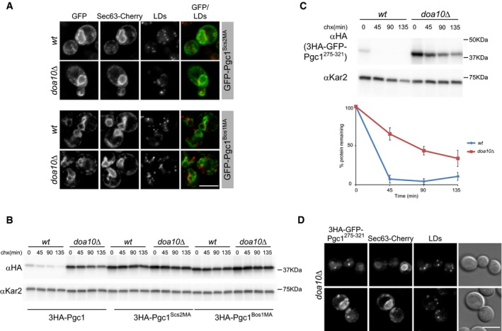
- Localization of GFP‐tagged Pgc1 derivatives in which the hydrophobic hairpin (aa275–321) was replaced by membrane anchor of Scs2 (Pgc1Scs2MA) or Bos1 (Pgc1Bos1MA). ER and LDs were visualized by expression of Sec63‐Cherry and MDH staining, respectively. Scale bar: 5 μm.
- The degradation of 3HA‐Pgc1 or the indicated chimeras in wt and doa10Δ cells was analyzed as in Fig 1A. The blot is representative of at least three independent experiments.
- The degradation of 3HA‐GFP‐Pgc1275–321 in wt and doa10Δ cells was analyzed as in Fig 1A. The graph shows the average of four independent experiments; error bars represent the standard deviation.
- Localization of 3HA‐GFP‐Pgc1275–321 in doa10Δ cells. The ER was visualized by expression of Sec63‐Cherry. Scale bar: 5 μm.
Source data are available online for this figure.