
Figure 6. A heterologous hydrophobic hairpin functions as a Doa10 degron.
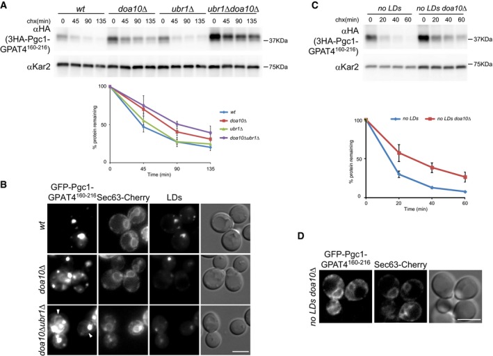
- The degradation of 3HA‐Pgc1‐GPAT160–216 containing GPAT4 hydrophobic hairpin (aa160–216) replacing the one of Pgc1 was analyzed in cells with the indicated genotype as in Fig 1A. The graph shows the average of three independent experiments; error bars represent the standard deviation.
- Localization of GFP‐Pgc1‐GPAT4160–216 in cells with the indicated genotype. Arrowheads indicate GFP‐Pgc1‐GPAT4160–216 labeling at the ER, marked with Sec63‐Cherry. LDs were visualized upon staining with MDH. Scale bar: 5 μm.
- The degradation of 3HA‐Pgc1‐GPAT160–216 in cells with the indicated genotype was analyzed as in Fig 1A. The graph shows the average of three independent experiments; error bars represent the standard deviation.
- Localization of GFP‐Pgc1‐GPAT160–216 in are1Δ are2Δ lro1Δ dga1Δ doa10Δ mutant (no LDs doa10Δ). The ER was visualized by expression of Sec63‐Cherry. Scale bar: 5 μm.
Source data are available online for this figure.