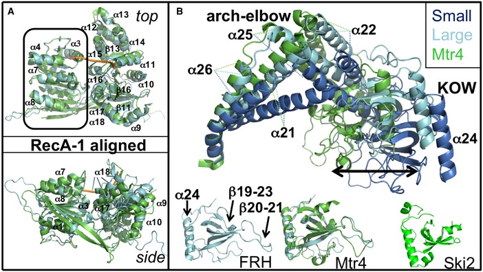
Figure 4. The RecA and arch domains of FRH compared to Mtr4.

- The two RecA domains are rotated relative to one another in FRH (cyan, large cell) compared to Mtr4 (green, RNA backbone in orange). The change in positioning of RecA‐2 with respect to RecA‐1 results in an overall displacement from the Mtr4 domain juxtaposition that ranges from 1.1 to 4.0 Å throughout RecA‐2 with the largest differences at the external helices.
- Comparison of arch domains conformations from Mtr4 (green) and FRH (large cell in cyan, small cell in marine) after superposition of the most N‐terminal helical region. The isolated KOW modules of FRH, Mtr4 (with FRH superimposed), and Ski2 have considerably different loop configurations (below).
