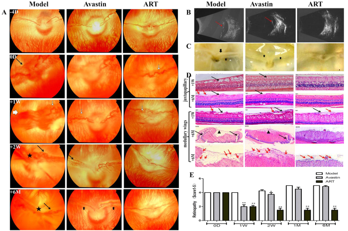
Figure 2. The effect of ART on rabbit retinal vascular disorders induced by VEGF165+bFGF.
(A–C) ART and Avastin significantly diminished vascular dilatation and tortuosity (white arrows in A) and NV (black arrows in A) at 1 week. However, NV and fluorescein leakage recurred in the Avastin group at 2 weeks and finally resulted in tractional detachment of the retina (thick black arrows in A and C, red arrows in B). The tractional retinal detachments occurred at 2 weeks in the model group (★ in A). No visible neovascular membrane occurred in the ART group for 6 months. (D) H&E staining of cross-sections of the medullary wings (medullary) and superior to the optic disc (juxtapapillary). ART and Avastin significantly inhibited retinal edema (black arrows) at 1 week compared to the model group but did not noticeably reduce epiretinal fibrovascular membranes (red arrows). After 6 months, epiretinal fibrosis (▲) and retinal folds (black arrows) appeared in the model and Avastin groups, along with vascular crowding, which was not found in the ART group. The inner retina was disorganized (black arrows) in the model group; this did not occur in the ART or Avastin groups. (E) Grade score for retinal vascular disorders. All fundus and FFA photographs of rabbits were evaluated independently and in a masked manner by two observers and were scored using the five grades of retinal NV. The grade scores were used for ANOVA analysis, and differences with a value of p < 0.05 were considered significant.

