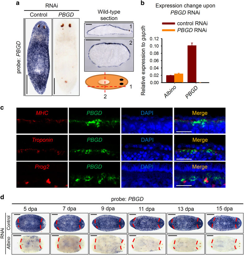
Figure 6.
PBGD labels pigment cells in planarian. (a) WISH and frozen section of PBGD in wild-type animals showing an epidermal-specific expression pattern. Scale bar: 200 μm. (b) Relative expression level to gapdh. Shown are averages of three independent experiments; error bars=s.d. (c) Double FISH for PBGD with mhc, troponin and prog2 in wild-type animals at dorsal body wall showing PBGD-positive cells lying in between muscle cells. Scale bar: 20 μm. Images are single confocal sections. (d) WISH showing expression patterns of PBGD in regenerating control or Albino RNAi worms. Red dashes indicate amputation sites. Scale bar: 200 μm.

