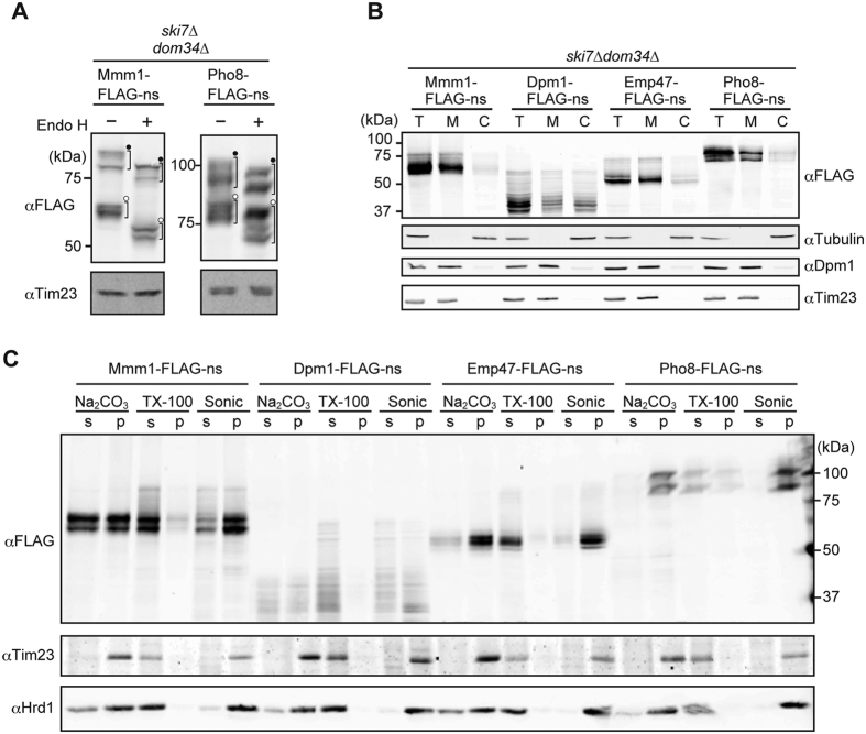
Figure 5. Cellular localization and membrane association of nonstop ER membrane proteins.
(A) Whole cell extracts prepared from ski7∆dom34∆ cells expressing Mmm1-FLAG-ns or Pho8-FLAG-ns from the GPD1 promoter were incubated with or without Endo H (500 unit/ml) at 37 °C for 40 min, and analyzed by SDS-PAGE and immunoblotting. Closed dots and open dots indicate tRNA-attached and tRNA-dissociated forms, respectively. (B) ski7∆dom34∆ cells expressing the indicated nonstop membrane proteins from the GPD1 promoter were fractionated by differential centrifugations. T, total; M, High-speed precipitate containing membrane fractions; (C) High-speed supernatant containing cytosolic fractions. (C) The membrane fractions prepared in (B) were treated with Na2CO3 at pH 9.4 or Triton X-100 (TX-100), or sonicated (sonic), and then subjected to ultracentrifugation. The resulting supernatant (s) and precipitate (p) were analyzed by SDS-PAGE and immunoblotting using the indicated antibodies.

