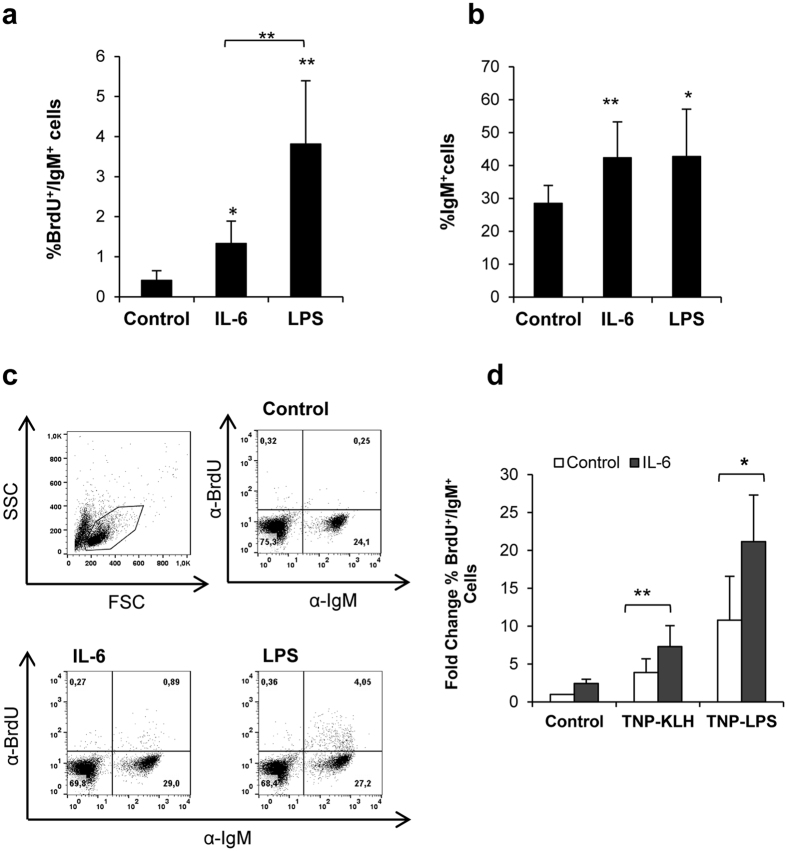
Figure 1. IL-6 and LPS elicit B cell proliferation and increased survival.
Spleen leukocytes were incubated with IL-6 (200 ng/ml) or LPS (100 μg/ml) for 3 days at 20 °C. After this time, BrdU was added to the cultures and incubated for a further 24 h. The percentage of proliferating (BrdU+) IgM+ B cells was determined as described in Material and Methods. (a) Percentage (mean + standard deviation) of proliferating IgM+ B cells (BrdU+/IgM+) after treatment with IL-6 and/or LPS (n = 6). (b) IgM+ B cell survival estimated as percentage of IgM+ cells (proliferating and non-proliferating cells) in cultures (mean + standard deviation) (n = 6). (c) Representative dot plots are shown. (d) Spleen leukocytes were stimulated with TNP-KLH (5 μg/ml) or TNP-LPS (5 μg/ml) in the presence or absence of IL-6 (200 ng/ml). The percentage of proliferating IgM+ cells was assessed as described above. Data are shown as the mean fold change relative to the control value for unstimulated controls + standard deviation (n = 5). Asterisks denote significant differences between cells treated with IL-6 or LPS and their corresponding controls and between IL-6 and LPS treated cells when indicated. *P < 0.05, **P < 0.01.

