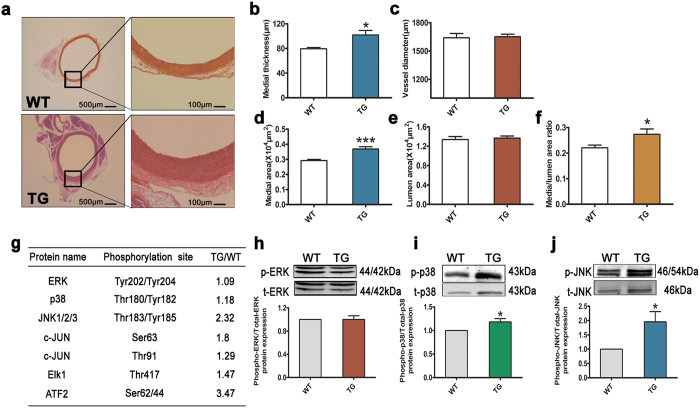
Figure 3. L3MBTL4 induces vascular remodeling via MAPK signaling pathway.
(a) Representative photomicrographs of hematoxylin-eosin staining in blood vessels from wild type rats (WTs) and transgenic rats (TGs) (n = 7 each group). (b–f) The media thickness, vessel diameter, media area, lumen area and media/lumen area ratio of aortas is quantified (n = 7 per group). Scale bars are 500 μm and 100 μm. (g) Changed phosphorylated proteins in the vessels of TGs compared to WTs are identified by phospho-antibody microarray. Listed are proteins in mitogen-activated protein kinases (MAPK) family. (h–j) Western Blot analysis validate the phosphorylation levels of extracellular signal-regulated kinase (ERK), p38MAPK and c-Jun N-terminal kinase (JNK) in the aortas from WTs and TGs; n = 7 per group for (h,j), n = 6 each group for (i). *p < 0.05, ***p < 0.001 compared to WT. All data represent mean ± s.e.m.

