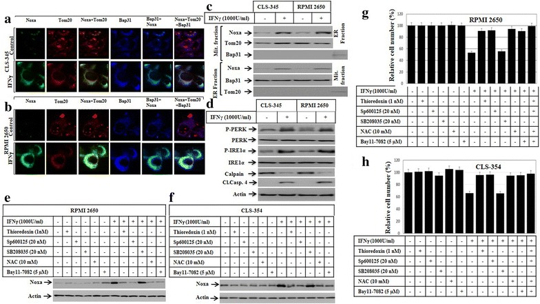
Fig. 6.

a Subcellular localization of Noxa protein to both mitochondria and endoplasmic reticulum (ER). Immune fluorescence: CLS-354 and RPMI 2650 cells were treated with IFNγ for 48 h before the staining with anti- Noxa, Tom20 (mitochondrial marker) and Bap31 (ER marker). The subcellular localization of Noxa (green) to mitochondria (red) and the overlay of Noxa with Tom20 staining demonstrates the localization of Noxa to mitochondria (yellow), when compared to control cells. b The subcellular localization of Noxa (green) to ER (blue) and the overlay of Noxa with Bap31 staining demonstrates the localization of Noxa to ER (turquoise), when compared to control cells. c Western blot analysis using mitochondrial fraction (Mit. fraction) and ER fraction from both CLS-354 and RPMI 2650 cells following the treatment with IFNγ for the indicated time periods. The detection of Noxa in mitochondrial and ER fractions of CLS-354 and RPMI 2650 cells after the exposure to IFNγ confirm the localization of Noxa protein to both mitochondria and ER. The purity of both mitochondrial and ER fractions was verified by the detection of the mitochondrial protein Tom20 in the mitochondrial fraction and the detection of Bap31 in ER fraction. d Western blot analysis demonstrates the phosphorylation of both PERK and IRE1α, calpain degradation, and cleavage of caspase-4 in response to the treatment of HNSCC cells with IFNγ. e Western blot analysis demonstrates the inhibition of IFNγ-induced expression of Noxa in RPMI 2650 in response to the pre-treatment with the inhibitors of the ASK1 (thioredoxin), JNK (SP600125), ROS (NAC) and NF-κB (Bay11-7082), but not with those of p38 (SB208035). f Western blot analysis demonstrates the inhibition of IFNγ-induced expression of Noxa in CLS-354 cells in response to the pre-treatment with the inhibitors of the ASK1 (thioredoxin), JNK (SP600125), ROS (NAC) and NF-κB (Bay11-7082), but not with those of p38 (SB208035). Actin was used as internal control for loading and transfer. Data are representative of three independent experiments. g MTT demonstrates the inhibition of IFNγ-induced death of RPMI 2650 cells in response the pre-treatment with the inhibitors of the ASK1 (thioredoxin), JNK (SP600125), ROS (NAC) and NF-κB (Bay11-7082), but not with those of p38 (SB208035). h MTT demonstrates the inhibition of IFNγ-induced death of CLS-354 cells in response to the pre-treatment with the inhibitors of the ASK1 (thioredoxin), JNK (SP600125), ROS (NAC) and NF-κB (Bay11-7082), but not with those of p38 (SB208035). The values are expressed as the mean ± SD of three independent experiments performed in duplicate. The Student’s t test was used for analysis
