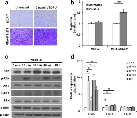
Fig. 4.

Migration and signaling pathways in VEGF-A-stimulated MDA-MB-231 cells. a Images of the crystal violet-stained cells that migrated horizontally in the trans-well migration assay. b The absorbance values of extracted crystal violet in migrated cells. Migration of the VEGF-A-treated MDA-MB-231 cells (~1.5-fold) was enhanced compared with that of the untreated control cells. c Representative image of western blotting of phosphorylated AKT, FAK and ERK and total AKT, FAK and ERK in MDA-MB-231 cells treated with VEGF-A. The levels of FAK and AKT phosphorylation were increased in the VEGF-A-treated MDA-MB-231 cells, but the level of ERK phosphorylation was similar between VEGF-A-treated MDA-MB-231 cells and the untreated cells. d Densitometric quantifications of phosphorylation levels of FAK, AKT, and ERK in the VEGF-A-treated cells relative to the untreated cells. β−actin was used as an internal reference. All experiments were performed at least in triplicate, and the values are the mean values ± standard deviation. *p < 0.05, **p < 0.001
